Aug 31 2009
In time for the 2009 Microscopy Conference in Graz, Austria, Carl Zeiss is presenting an integrated solution for correlative microscopy in materials analysis. Core elements of the link between light microscopy and scanning electron microscopy (SEM) are the sample holder and adapter, as well as software for the fast, automated relocation and examination of one and the same region of interest in the light microscope as well as in the SEM. This solution, called "Shuttle + Find," can be applied in both materials science as well as in many industrial routine procedures. Shuttle + Find will be widely available upon completion of the first applications that are being developed jointly with early customers. For the time being the solution is not suited for biological applications.

While light microscope images are based on reflection, or transmission, or fluorescence, the scanning electron microscope offers - in addition to the more than two orders of magnitude higher resolution - a variety of more in-depth analysis possibilities. These options primarily include the morphological characterization of the sample and the element analysis using X-ray spectroscopy (EDX). Initially, samples will be examined under a light microscope deploying various contrasting methods in bright field, dark field or using polarized light or interference. Interesting areas of the sample are then further examined in detail under the scanning electron microscope.
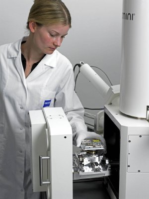
The "Shuttle + Find" solution from Carl Zeiss for correlative microscopy in materials analysis is an easy-to-use interface. It connects upright and inverted light microscopes of type SteREO Discovery, Axio Imager and Axio Observer, featuring a motorized stage, with all ZEISS scanning electron microscopes such as EVO, SIGMA, SUPRA, ULTRA and MERLIN, as well as the CrossBeam (FIB-SEM) workstations AURIGA, NVision and NEON. Within a few minutes, it is possible to transfer a sample from one system to the next. The sample can also be sent to another lab equipped with an SEM. In the SEM, the areas of interest of the sample examined are automatically relocated in a matter of seconds. In addition to numerous analyses which are based on separate images from light and electron microscopes the images of the two different instrument platforms can be precisely overlaid and correlated to X-ray maps to deliver further information.
Dr. Thomas Albrecht, Head of Product Management at Carl Zeiss SMT´s Nano Technology Systems division emphasizes: "As the only provider in the world to develop, produce and sell both light and electron microscopes, Carl Zeiss is predestined to develop solutions for correlative microscopy. With the interface presented today, we are taking a step which enables two largely separate worlds to come together. Our ´Shuttle + Find´ solution really means Bridging the Micro and Nano World".