Feb 21 2017
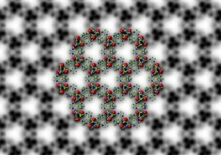
 Symmetry-imposed and lattice-averaged HRTEM image of the metal-organic framework ZIF-8 (black and white) with a structural model overlaid to show the position of the zinc ions and organic ligands (in color). CREDIT: 2017 KAUST Ivan D. Gromicho.
The atomic structure of metal-organic frameworks (MOFs) - three-dimensional structures formed of metal ions connected through organic ligands - can be analyzed by using high-sensitivity electron cameras.
A research team from the King Abdullah University of Science and Technology (KAUST) has developed an innovative technique to carry out fine-scale imaging of MOF. The ability of the MOF to be fabricated to have large void spaces, or porosity, within their frameworks as well as accurate pore sizes of molecular dimensions renders them highly applicable for gas storage and separation.
Conventionally, structures with atomic resolution are visualized using high-resolution transmission electron microscopy (HRTEM). Yet, this technique is inappropriate for the visualization of MOFs as the electron beams damage the structures of the MOF.
To thoroughly understand the performance of metal-organic frameworks in various applications, we need to know their structures at the atomic level because their macroscopic behavior is determined by their microscopic structure.
Yu Han, Associate Professor of Chemical Science at KAUST
Visualization of these structures can help researchers to unearth significant hints related to the way in which the MOF self-assemble to form their characteristic pores.
Various members from the Advanced Membranes and Porous Materials Center at KAUST, including Yihan Zhu, Han’s research scientist and first author of the paper; Zhiping Lai, Associate Professor of Chemical and Biological Engineering; and Ingo Pinnau, Professor of Chemical and Biological Engineering as well as Director of the Center, worked in collaboration with the Imaging and Characterization Core Lab at KAUST and with collaborators from Gatan, Lawrence Berkeley National Laboratory and others in China. This collaboration has led to the use of HRTEM in tandem with state-of-the-art direct-detection electron-counting cameras.
The researchers exploited the high sensitivity of the detectors to obtain images with low-enough electron doses which do not destroy the structure of the MOF, allowing the researchers to generate high-resolution images of the atomic structures of MOF.
The researchers applied their technique to ZIF-8, a MOF comprising of zinc ions connected through organic 2-methylimidazole linkers. The team was successful in imaging its structure at a resolution of 0.21 nm, which is far greater for imaging the individual columns of organic linkers and zinc atoms, where 1 nm is one billionth of 1 m.
This assisted the research team to unearth the interfacial and surface structures of ZIF-8 crystals.
The results unraveled that porosity generated at the interfaces of ZIF-8 crystals is different from the intrinsic porosity of ZIF-8, which influences how gas molecules transport in ZIF-8 crystals.
Yu Han, Associate Professor of Chemical Science at KAUST