Jan 21 2019
Scientists at MIT have devised a method for achieving a drastic improvement in the sensitivity of nuclear magnetic resonance spectroscopy (NMR)—a technique adopted for analyzing the structure and composition of different types of molecules, such as proteins associated with Alzheimer’s and other diseases.
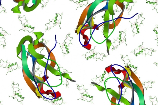
 MIT chemists have enhanced the resolution of nuclear magnetic resonance (NMR), which should allow more rapid and detailed study of proteins such as the beta amyloid protein that accumulates in the brains of Alzheimer’s patients. (Image credit: MIT News)
It is considered that this innovative method will enable researchers to study structures in just minutes, which would have previously taken years to figure out, stated Robert Griffin, the Arthur Amos Noyes Professor of Chemistry. The new strategy, which is dependent on short pulses of microwave power, could enable scientists to determine the structures of various complex proteins that have been hard to analyze to date.
This technique should open extensive new areas of chemical, biological, materials, and medical science which are presently inaccessible.
Robert Griffin, Study Senior Author, Arthur Amos Noyes Professor of Chemistry, MIT
The lead author of the study, which has been published in the Sciences Advances journal on January 18th, 2019, is MIT postdoc Kong Ooi Tan. The other authors of the study are former MIT postdocs Chen Yang and Guinevere Mathies, and Ralph Weber of Bruker BioSpin Corporation.
Improved Sensitivity
In conventional NMR, the magnetic properties of atomic nuclei are exploited to uncover the structures of the molecules that contain those nuclei. NMR uses a strong magnetic field with the ability to interact with the nuclear spins of hydrogen and other isotopically labeled atoms, such as nitrogen or carbon, to measure a property called chemical shift of these nuclei. Those shifts are specific for each atom and thus act as fingerprints, which can be further used to unravel the way these atoms are linked.
NMR’s sensitivity is dependent on the polarization of atoms—a measure of the variation between the number of “up” and “down” nuclear spins in each spin ensemble. The sensitivity that can be achieved is directly proportional to the extent of polarization. As a rule, a stronger magnetic field of up to 35 Tesla is applied by scientists to enhance the polarization of their samples.
Another strategy, which has been under development over the past 25 years by Griffin and Richard Temkin of MIT’s Plasma Science and Fusion Center, further increases the polarization with the help of a method known as dynamic nuclear polarization (DNP). In this method, the polarization is transferred from the unpaired electrons of free radicals to carbon, hydrogen, phosphorus, or nitrogen nuclei in the sample under analysis. This improves the polarization and renders it easier to reveal the structural features of the molecule.
In general, DNP is carried out through continuous irradiation of the sample with high-frequency microwaves, which is achieved with the help of a device known as a gyrotron. Thus, NMR sensitivity is enhanced by nearly 100-fold. Yet, this technique necessitates immense power and does not work well at higher magnetic fields that could enable resolution improvements that are even greater.
In order to solve this issue, the MIT researchers devised a process for delivering short pulses of microwave radiation, rather than continuous microwave exposure. They delivered these pulses at a particular frequency and could improve polarization by a factor of up to 200. Although this is analogous to the enhancement accomplished using conventional DNP, it necessitates just 7% of the power, and in contrast to conventional DNP, it can be executed at higher magnetic fields.
We can transfer the polarization in a very efficient way, through efficient use of microwave irradiation. With continuous-wave irradiation, you just blast microwave power, and you have no control over phases or pulse length.
Kong Ooi Tan, Study Lead Author, Postdoc, MIT
Time-Saving
According to the team, this enhancement in sensitivity will enable the analysis of samples that would have earlier have taken almost 110 years to study in a single day. The researchers demonstrated the technology in the Sciences Advances paper by using it to study standard test molecules, for example, a glycerol-water mixture; however, currently, they intend to use it on highly complex molecules.
One important area of interest is the amyloid beta protein that gets accumulated in the brains of Alzheimer’s patients. The team also intends to analyze a wide range of membrane-bound proteins, for instance, ion channels and rhodopsins, which are light-sensitive proteins contained in bacterial membranes and in the human retina. Thanks to its higher sensitivity, this technique can extract useful data from considerably smaller sample size, probably rendering it easier to analyze proteins that are hard to obtain in large quantities.
The National Institutes of Biomedical Imaging and Bioengineering, the Swiss National Science Foundation, and the German Research Foundation funded the study.