The ZEISS Xradia Context is a non-destructive 3D X-Ray micro-computed tomography instrument with a broad field of view. Users can photograph tall, heavy (25 kg) and large samples in their complete 3D environment, as well as tiny samples with excellent resolution and detail, using a powerful stage and adjustable software-controlled source/detector configuration.
- Use non-destructive failure analysis to find internal flaws in user sample or workpiece without having to cut it
- Connect to the ZEISS correlative microscopy environment and use nondestructive 3D imaging to find regions of interest for high-resolution 2D or 3D electron microscopy imaging
- Porosity, fractures, inclusions, flaws and various phases are examples of performance-defining heterogeneities in complex materials that can be characterized and quantified
- Get 3D data on whole electrical components, massive raw materials samples or biological specimens
- Ex situ or in situ sample modifications are used to conduct 4D evolutionary investigations
Highlights
Full Context 3D Imaging
- With tiny samples, increase geometric magnification to locate and describe micron-scale features with excellent contrast and clarity
- With picture quality, users may go up to 10X quicker
- Quickly mount and align the specimens, or upgrade to an Autoloader for automated specimen collection and continuous scanning of up to 14 samples
- With a high pixel density detector of six megapixels, users can resolve small details in their complete 3D context, even in a rather large image volume
- Benefit from a simplified collection approach that allows for quick exposure periods and data restoration

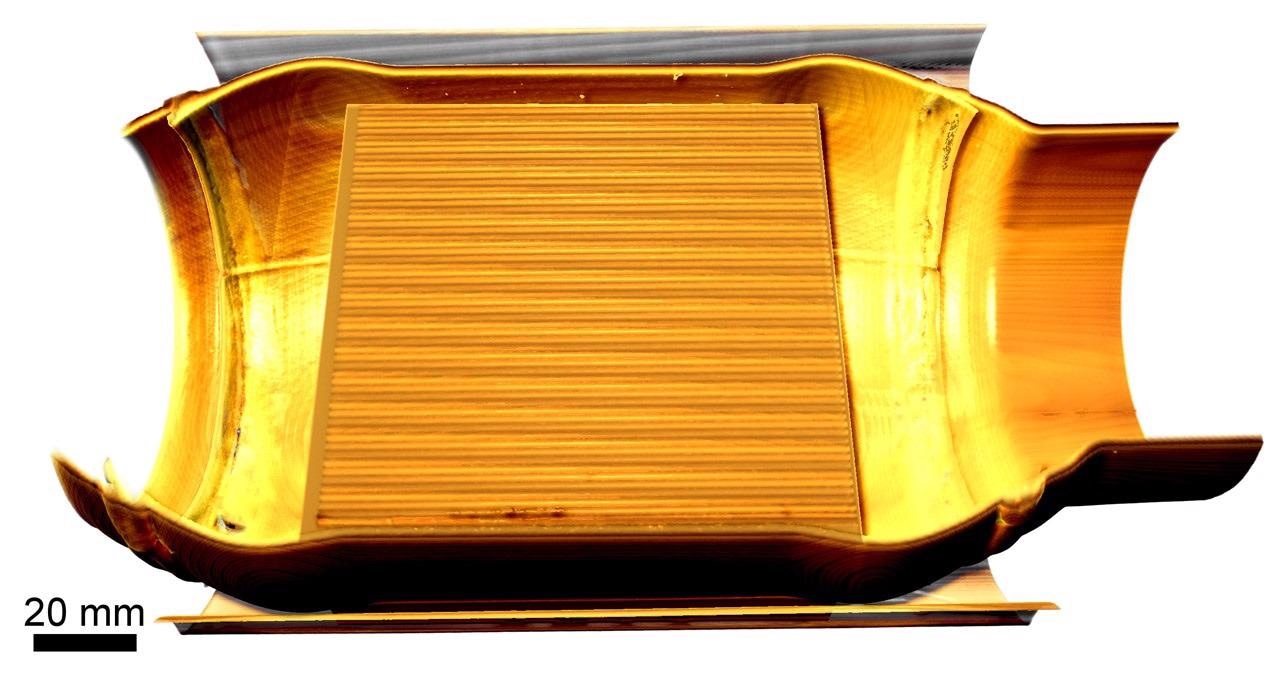
Virtual cutaway view of the interior of an intact catalytic converter. Image Credit: Carl Zeiss Microscopy GmbH
Based on the Proven Xradia Platform
- Take advantage of a system designed to ensure stability, as well as developments in high-resolution, high-quality data capture and reconstruction
- Conduct 4D experiments to determine how the microstructure of materials varies under different situations
- The Scout-and-Scan control system is simple to use and provides an effective working environment
- As it is based on the same technology as the Xradia Versa series, the Xradia context microCT benefits from years of research

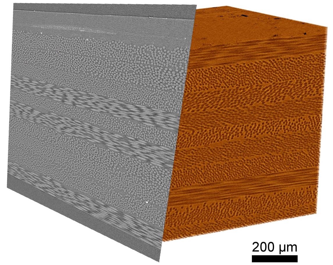
3D rendering and clipping plane of a composite sample with carbon and glass fibers. Image Credit: Carl Zeiss Microscopy GmbH
Convertible to X-Ray Microscopy (XRM)
- This enables users to use 3D tomographic imaging that can scale to meet user needs. The only microCT that can be converted to a ZEISS Xradia 5XX Versa 3D X-Ray microscope at any time is the Xradia Context microCT (XRM)
- For more flexibility and performance in spatial resolution, improved contrast and acquisition methods, add FPX (flat panel extension)
- To examine material evolution in 4D, use the in situ Interface Kit and load stages
- Users’ imaging instruments should grow in parallel with customer needs: Xradia Context is now part of the ZEISS X-Ray imaging portfolio, allowing ZEISS to continue to expand the capabilities and usefulness of its field systems

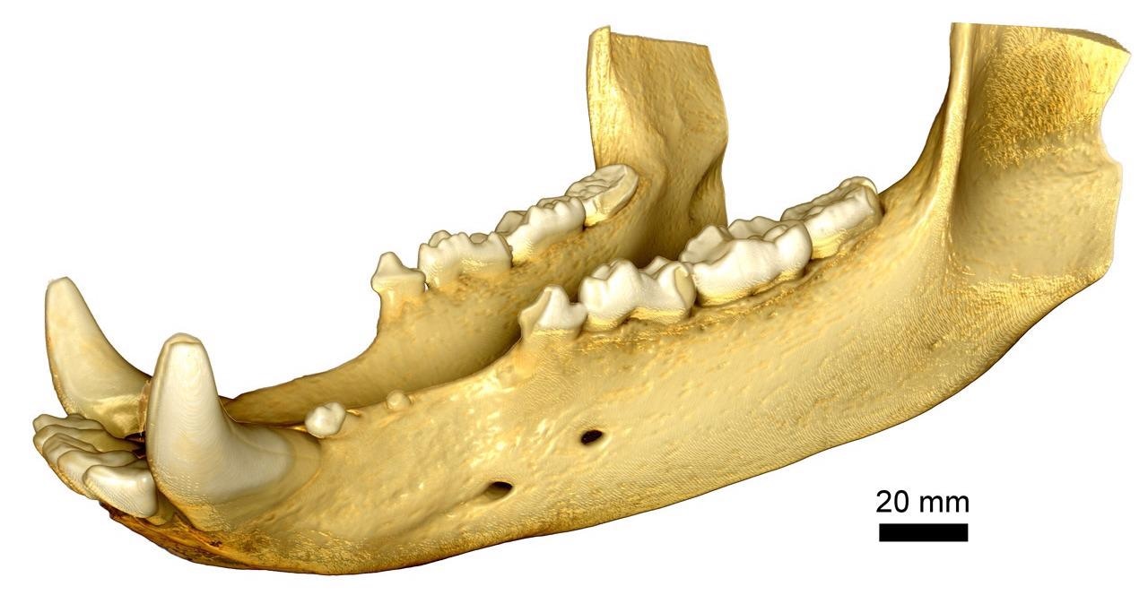
Full field of view imaging of a bear jaw. Image Credit: Carl Zeiss Microscopy GmbH