May 23 2019
A wish for a simpler, economical way to conduct standard laboratory tests for medical diagnoses and to escape “washing the dishes” led University of Connecticut scientists to create a new technology that decreases cost and time.
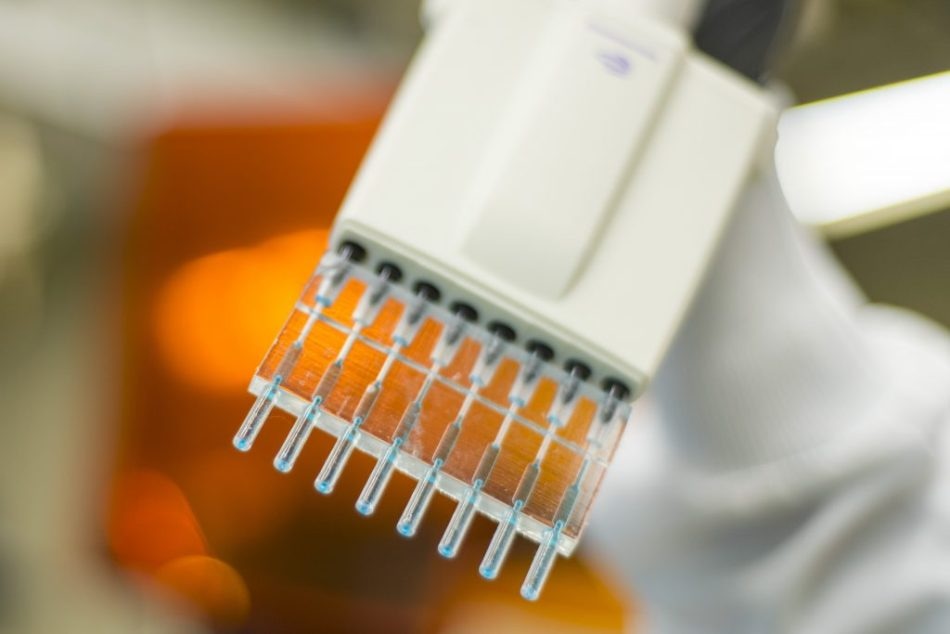
 Mohamed Sharafeldin, holds a unique pipette tip created with a 3D printer. (Sean Flynn/UConn Photo)
Their pipette-based technology could also help make specific medical testing available in remote or rural areas where traditional techniques might otherwise be excessively expensive and difficult to conduct.
The 3D-printed pipette-tip test designed by the team leverages what “has long been the gold standard for measuring proteins, pathogens, antibodies and other biomolecules in complex matrices,” they say. The technique still uses the enzyme-linked immunosorbent assay, also called ELISA, but through a different path. They have described their findings in a paper now published online in Analytical Chemistry.
For three decades or more, ELISA has been employed to test blood, cells, and other biological samples for all illnesses from certain cancers to HIV, from pernicious anemia to Lyme disease.
Traditional ELISA tests are carried out on plates having 96 micro-wells; each well acts as an independent testing chamber where samples can be integrated with different agents that will then react with the sample, usually by changing color. Technicians can then examine whether a sample shows indicators of a specific disease or condition based on the intensity of the color generated during the reaction.
While successful and accurate, the equipment used to work ELISA is costly – typically costing thousands of dollars to set up in a lab – and necessitates specialized training to carry out testing, as inappropriate methods can lead to flawed results. The agents used in the real tests – typically different forms of antibodies – can be costly as well.
Similar to many research laboratories, James Rusling’s chemistry lab where research assistant Mohamed Sharafeldin and his primary collaborator, Karteek Kadimisetty ’18 Ph.D., conducted their study, does not have an automated ELISA washing machine, meaning that plates being used for tests must be washed by hand – a laborious and demanding process.
“The ELISA washing techniques take forever,” said Sharafeldin, who is presently working toward his doctorate in chemistry. “It’s very tough, especially in a lab like ours. We don’t have those kind of fancy washing machines.”
When Kadimisetty was running ELISA at some point, he stated, “I wish doing ELISA was as simple as pipetting.” That casual comment was the impetus for what ensued: a design for a 3D-printed adapter for frequently used pipettes that could work an ELISA test directly in the pipette tip, minus the need for a traditional ELISA plate and the costly equipment that goes with it.
Each single-use pipette tip signifies one micro-well on an ELISA plate; the scientists also designed a multi-tipped model that allows for eight tips to be pipetted simultaneously. The tips fit tightly onto the majority of pipettes used in laboratory settings, making fluid handling a lot easier than with the typical ELISA plate.
We didn’t want to make a big change in the traditional ELISA; we just made engineered, controlled changes. So, the basics are the same. We use the same antibodies at the same concentrations that they use with conventional or traditional ELISA, so we are using the same protocols. Anything that can be run by normal ELISA can be run by this, with the advantage of being less expensive, much faster and accessible.
Mohamed Sharafeldin, Research Assistant, James Rusling’s Chemistry Lab, University of Connecticut
The scientists tested the pipette tips on samples from prostate cancer patients and learned not only were the test results from the tips as exact as ELISA tests, but also they were able to carry out the tests with one-tenth of the amount of testing agent – considerably decreasing the overall cost of the test – and at a fraction of the time. Tests performed by various users with varying degrees of skill ultimately showed the same results.
Traditional ELISA plate micro-wells contain 400 microliters of fluids each, but the reactions required to measure test results only happen on the plastic walls of the well. While the 3D-printed ELISA tips can contain just 50 microliters, the reservoir’s design inside the tip greatly increases the surface area where reactions take place, allowing the scientists to use a lot less of the expensive antibodies used to perform the test, and considerably reducing the time taken to process the test and read the results.
“Here we have a chamber where the reaction happens at all points,” Sharafeldin said, talking about the pipette tip design. “This reduces the time of the assay, which is an important thing, because the ELISA assay takes from five to eight hours to run. This one can be run in 90 minutes.”
The pipette tips also do not require a costly or sophisticated plate reader to establish test results, as ELISA tests demand. In the experiments with the prostate cancer samples, the pipette tip results were correctly read by clicking a cell phone photo and using a free app that computes color intensities in the image.
The advantage, Sharafeldin said, is that the user performing the test with the pipette tips does not have to be skilled; they merely need basic pipetting instructions, then to click a photograph and transmit it to a technician who could remotely read the results to help come with up a diagnosis – potentially offering new, lower-cost testing options in rural or isolated areas where setting up a traditional ELISA lab would prove difficult and costly.
While further sample testing is required, Sharafeldin is confident about the future prospect for the pipette tip design to lower costs. He is also coordinating with engineers to build an automated, vacuum-assisted pipette that would further simplify the use of the pipette tips and the conducting of ELISA tests, and would be available for considerably less cost than traditional ELISA equipment.
Besides Sharafeldin, Kadimisetty, and Rusling, collaborators include Ketki R. Bhalerao, Itti Bist, Abby Jones, Tianqi Chen; and Norman H. Lee, professor of pharmacology and physiology at George Washington University.
The project was aided by a UConn Academic Plan Grant and partly supported by Grant No. EB016707 from the National Institute of Biomedical Imaging and Bioengineering (NIBIB), NIH.