.jpg) By Susha Cheriyedath, M.Sc.Reviewed by Skyla BailyNov 17 2021
By Susha Cheriyedath, M.Sc.Reviewed by Skyla BailyNov 17 2021The spatial resolution of anisotropic charge distributions on an atom has been an unsolved scientific challenge. A group of researchers from the Czech Republic has demonstrated that a properly actualized kelvin probe force microscopy can image the anisotropic charge of the σ-hole and quadrupolar charge of a carbon monoxide molecule. This experiment has been published in the journal SCIENCE.

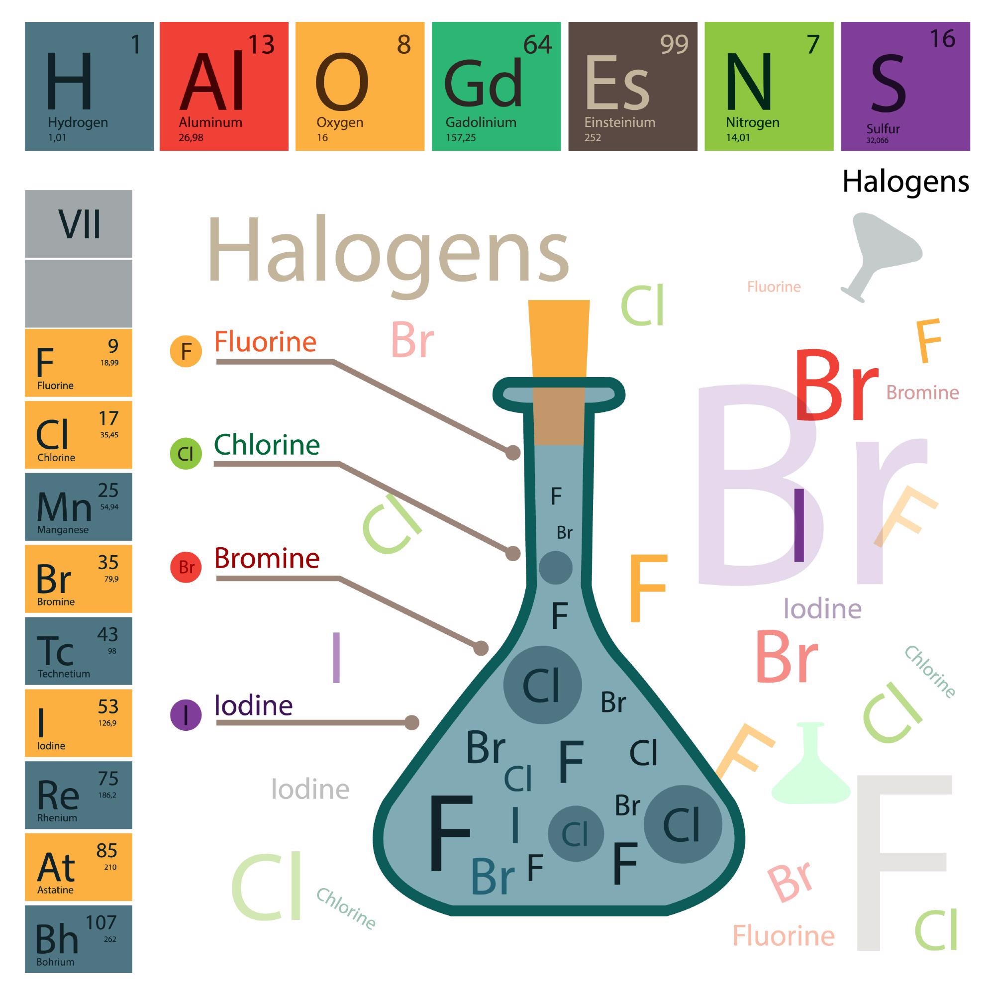
Study: Real-space imaging of anisotropic charge of σ-hole by means of Kelvin probe force microscopy, Image Credit: Yaruna/Shutterstock.com
Why Halogen-Bonding is Peculiar
The atomic arrangement of two adjacent halogens or a pair of halogen atoms and electron-donating atoms like oxygen, nitrogen, and sulfur, have unusual molecular structures. It represents a long-pending enigma in supramolecular chemistry.
Theoretically, close contacts between both halogens and electron donor motif atoms should cause a highly repulsive electrostatic reaction because both of them carry a negative charge. However, such atoms are frequently found to form intermolecular bonds, known as halogen bonds, that stabilize the molecular structure.
What are the Prevalent Theories on Halogen-Bonding?
Some researchers have shown that the formation of a covalent bond between certain halogen atoms such as bromine, chlorine, and iodine and a more electronegative atom like carbon forms a σ-hole that has an anisotropic charge distribution on the halogen atom.
Hence, a physically observable electrostatic potential on the halogen atom is not uniform but possesses an electropositive distal to covalently bond a carbon atom surrounded by an electronegative belt.
As a result, halogen bonding is attributed to attractive electrostatic forces between a halogen’s electropositive σ-hole, an electronegative atom with a negative charge, and/or an electronegative belt of the other halogen.
The stability of the σ-hole bonding in both hydrogen-bonded molecules and non-covalent molecules is comparable, and is due to the electrostatic interaction. However, this scenario is valid for H-bonded molecules. In the case of halogen-bonded systems, the dispersion interaction is because of close contact between two heavy atoms with a high polarizability.
The understanding of halogen bonding was later generalized to a σ-hole bonding concept. Particularly, the tetrel (group 14), pnicogen (group 15), chalcogen (group 16), halogen (group 17), and aerogen bonding (group 18) were formularized according to the name of the electronegative atom bearing the positive σ-hole.
Scientists become first to observe an inhomogeneous electron charge distribution on an atom
Video Credit: IOCB Prague/Youtube.com
The evidence of the σ-hole was established indirectly with crystal structures of compounds containing σ-hole donors and electron acceptors or quantum calculations. However, a direct representation of this entity allowing for the resolution of its peculiar shape has been absent.
What Does the Research Say?
The imaging of anisotropic atomic charge is a challenge for current experimental techniques, which include scanning probe microscopy (SPM), electron microscopy (EM), and diffraction methods due to the anisotropic distribution of the atomic charge on a halogen atom.
The team applied a technique called kelvin probe force microscopy (KPFM) in which the imaging mechanism relies on the electrostatic force to visualize the anisotropic charge distribution on a halogen atom with a sub-angstrom spatial resolution. KPFM was carried out under ultrahigh vacuum (UHV) conditions. The real-space visualization of the σ-hole gave undocumented spatial resolution.
What is Kelvin Probe Force Microscopy?
KPFM is a type of SPM technique that provides a real-space atomic resolution of surfaces. In the KPFM technique, the variation of the frequency shift of an oscillating probe on applied bias voltage with the quadratic form is recorded.
The vertex of the Kelvin parabola determines the difference between work functions of the tip and specimen, called the contact potential difference VCPD. Moreover, the spatial variation of the contact potential difference VCPD across the surface allows the mapping of local variation of the surface dipole on the sample (VLCPD).
The KPFM technique under ultra-high vacuum conditions gives true atomic resolution on surfaces, to image intramolecular charge distribution, to resolve bond polarity, to control single-electron charge states, or to discriminate charge.
There are two important components of this force: the static charge on the sample and the interaction between the polarized charge on the apex drt, which is linearly proportional to the applied bias voltage. The second term consists of the electrostatic interaction between the polarized charge on the sample and the static charge on the tip.
Finally, these two components cause local variation of the contact potential difference VLCPD.
Reference:
B. Mallada, A. Gallardo, M. Lamanec, B. De la torre, V. Spirko, P. Hobza, and P. Jelinek. Real-space imaging of anisotropic charge of σ-hole by means of Kelvin probe force microscopy, Science, 2021, 374, 6569, 863-867. DOI: https://www.science.org/doi/10.1126/science.abk1479
Disclaimer: The views expressed here are those of the author expressed in their private capacity and do not necessarily represent the views of AZoM.com Limited T/A AZoNetwork the owner and operator of this website. This disclaimer forms part of the Terms and conditions of use of this website.