The XGT-9000 X-ray analytical microscope from HORIBA Scientific is an evolution of μXRF. The microscope offers a combination of enhanced sensitivity and new imaging technology to realize the high-speed analysis of foreign materials using a single unit.
Features
- Easy analysis operation without the need for preparation and non-destructive analysis
- Complete with a range of image analysis software provided
- Measurement points can be rapidly accessed via high precision optical observation, even in the microscopic range
XGT-9000
Video Credit: HORIBA Scientific
Clear and High-Speed Image Mapping
- Reduced analysis time ensures efficient measurement work
- X-ray images with very limited noise enable even clearer observation

Image Credit: HORIBA Scientific
Clear Optical Image Observation and Coaxial X-Ray Irradiation
- Fitted with three types of illumination: transmitted, around and coaxial. Integrating coaxial and around illuminations allows clear observation of samples with regions that are mirrored, non-uniform, etc.
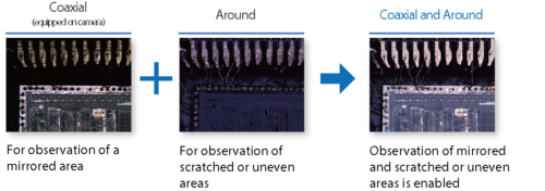
Image Credit: HORIBA Scientific
- Coaxial, perpendicular X-ray exposure and optical image observation avoid misalignment of the measurement position of samples from becoming uneven.
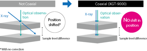
Image Credit: HORIBA Scientific
Employing of Imaging Technology Based on Raman Imaging Technique
A specific element is highlighted as a characteristic point, thus streamlining the detection of obstacles, foreign material, etc.
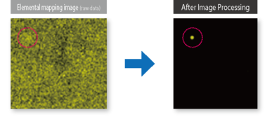
Image Credit: HORIBA Scientific
A New Solution in Foreign Material Analysis
The XGT-9000 enables foreign material to be rapidly detected via high-speed screening and highlighting by imaging processing. The high-resolution X-ray beam allows an elaborate analysis of elements constituting the foreign material. This sequence of foreign material analyses can be done with the help of a single unit, to the level of several tens of micrometers.
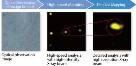
Image Credit: HORIBA Scientific
Applications
Analysis of Foreign Material in Films
The XGT-9000 supports visual confirmation, detection, and analysis of even hard-to-detect foreign materials while verifying optical observation images with high resolution following elemental mapping screening.
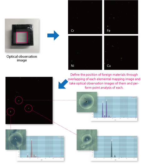
Image Credit: HORIBA Scientific
Analysis of Hydrous Samples
The XGT-9000 allows the measurement of hydrous samples and detection of foreign materials with the help of image processing.
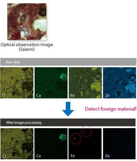
Image Credit: HORIBA Scientific
Specifications
Source: HORIBA Scientific
| Model |
XGT-9000 |
XGT-9000SL |
| Basic information |
| Instrument |
X-ray fluorescence analytical microscope |
| Sample type |
Solids, Liquids, Particles |
| Detectable elements |
C* – Am *with optional light elements detector (F – Am with standard detector) |
| Available chamber size |
450(W) x 500(D) x 80(H) |
1030(W) x 950(D) x 500(H) |
| Maximum sample size |
300(W) x 250(D) x 80(H) |
500(W) x 500(D) x 500(H) |
| Maximum mass of a sample |
1 kg |
10 kg |
| Optical observation |
Two high-resolution cameras with an objective lens |
| Optical design |
Vertical-Coaxial X-ray and Optical observation |
Sample illumination/
observation |
Top, Bottom, Side illuminations/Bright and Dark fields |
| X-ray tube |
| Power |
50 W |
| Voltage |
Up to 50 kV |
| Current |
Up to 1 mA |
| Target material |
Rh |
| X-ray optics |
| Number of probes |
Up to 4 |
Primary X-ray filters for
spectrum optimization |
5 positions |
| Detectors |
| X-ray Fluorescence detector |
Silicon Drift Detector (SDD) |
| Transmission detector |
NaI(Tl) |
| Mapping analysis |
| Mapping area |
100 mm x 100 mm |
350 mm x 350 mm |
| Step size |
2 mm |
4 mm |
| Operating mode |
| Sample environment |
Full vacuum / Partial vacuum /
Ambient condition / He purged condition(Optional) |
Partial vacuum / Ambient condition / He purged condition (optional)*
*He purge condition is necessary to detect down to carbon and fluorine for both detectors. |