The Helios 5 Hydra DualBeam (plasma focused ion beam scanning electron microscope, PFIB-SEM) from Thermo Scientific has the ability to provide four different ion species as the primary beam.
This enables users to select the ions that offer the best outcomes for their samples and use cases, like transmission electron microscopy (TEM) and scanning transmission electron microscopy (STEM) sample preparation and 3D materials characterization.
It is simple for users to switch between argon, oxygen, nitrogen and xenon in just 10 minutes without sacrificing performance. This unparalleled flexibility considerably extends the possible application space of PFIB and allows the study of ion-sample interactions to optimize present use cases.
The Helios 5 Hydra DualBeam integrates the new and inventive multi-ion-species plasma FIB (PFIB) column with the monochromated Thermo Scientific Elstar UC+ SEM Column to offer the latest focused ion- and electron-beam performance. Intuitive software, an unparalleled level of automation and user-friendliness enable observation and analysis of appropriate subsurface volumes.
Helios 5 Hydra DualBeam for Materials Science
High-quality sample preparation tends to be crucial for successful high-resolution material analysis with TEM or STEM. It asls is considered to be one of the most difficult and laborious tasks performed in materials characterization laboratories.
Traditional techniques that are utilized to make ultra-thin samples needed for S/TEM are slow and can need hours, or even days, of effort by highly trained personnel. This is complicated further by the range of various materials and the need for site-specific information. Plasma FIB, linked with proprietary and innovative software for automation and user-friendliness, resolves many of these difficulties.
Xenon plasma FIB, for instance, is considered to be the standard for gallium-free sample preparation, which is essential for sensitive samples like aluminum-containing materials.
The addition of fast switching between all four ion beam species of the Helios 5 Hydra DualBeam goes even further to fulfill the requirements of each of the users’ individual materials. This enables users to determine the perfect conditions to make the highest quality samples possible.
Helios 5 Hydra DualBeam for Life Sciences
The Helios 5 Hydra DualBeam opens new and undiscovered applications in life sciences by integrating high-resolution FIB-SEM tomography and high-throughput plasma technology. Users can make use of the optimal ion beam for every sample as a result of its advanced inductively coupled plasma (ICP) FIB along with four ion species.
Supreme surface quality has been achieved for every sample, irrespective of the preparation method or resin type. Besides higher milling throughput and sample compatibility, a new software suite eases persistent, high-quality, Ga-free, large-volume acquisition and 3D data analysis.
For life science research, the Helios Hydra DualBeam provides:
- Milling that can be performed without artifacts, irrespective of the sample preparation method
- Quick and efficient milling available to access 10× greater volumes compared to Ga-FIB milling
- Nanometer-thick serial sectioning of huge horizontal surfaces (up to 1 mm in diameter) with Spin Mill
- An adaptable solution for multi-user environment, compatible with an extensive range of samples and experimental questions
- Completely automated 3D data collection, higher throughput when integrated with Auto Slice and View Software

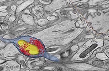
3D reconstruction of a mouse hippocampal organotypic slice culture. Image Credit: S. Watanabe, John Hopkins University
Key Features
Large Application Space
Largest application space available with special ion source delivering four fast, switchable ion species: N, O, Ar and Xe.
High Throughput and Quality
High-quality, high-throughput and statistically appropriate 3D characterization, cross-sectioning and micromachining can be performed with the help of a next-generation 2.5 μA plasma FIB column.
Advanced Automation
Quickest and simplest, automated, multi-site in situ and ex situ TEM sample preparation and cross-sectioning with the help of optional AutoTEM 5 Software.
High-Quality Sample Preparation
High-quality gallium-free TEM and APT sample preparation can be performed with xenon, argon or oxygen with the help of the new PFIB column allowing 500 V final polishing and delivering excellent performance at all operating conditions.
Short Time to Nanoscale
The shortest time to nanoscale data ensured for users with any experience level with FLASH and SmartAlign technologies.
Complete Sample Information
The most complete sample data is available with sharp, polished and charge-free contrast realized using up to six combined in-column and below-the-lens detectors
Electron and Ion Beam-Induced Deposition and Etching
The latest abilities are offered for electron and ion beam-induced deposition and etching on DualBeam systems with optional Thermo Scientific MultiChem or GIS Gas Delivery Systems.
Precise Sample Navigation
Customized to separate application requirements as a result of the high stability and precision of the 150 mm Piezo stage or the adaptable 110 mm stage and optional in-chamber Thermo Scientific Nav-Cam Camera.
Artifact-Free Imaging
This feature depends on combined sample cleanliness management and dedicated imaging modes like DCFI and SmartScan Modes
Specifications
Source: Thermo Fisher Scientific – Electron Microscopy Solutions
| |
Helios 5 Hydra CX DualBeam |
Helios 5 Hydra UX DualBeam |
| Electron beam resolution |
- At optimum WD:
- 0.7 nm at 1 kV
- 1.0 nm at 500 V (ICD)
- At coincident point:
- 0.6 nm at 15 kV
- 1.2 nm at 1 kV
|
| Electron beam parameter space |
- Electron beam current range: 0.8 pA to 100 nA at all accelerating voltages
- Accelerating voltage range: 350 V – 30 kV
- Landing energy range: 20* eV – 30 keV
- Maximum horizontal field width: 2.3 mm at 4 mm WD
|
| Ion optics |
High-performance PFIB column with unique inductively coupled plasma (ICP) source supporting four ion species with fast switching capability
- Ion species (primary ion beam): Xe, Ar, O, N
- Switching time <10 minutes, only software operation
- Ion beam current range: 1.5 pA to 2.5 µA
- Accelerating voltage range: 500V – 30 kV
- Maximum horizontal field width: 0.9 mm at beam coincidence point
Xe Ion beam resolution at coincident point
- <20 nm at 30 kV using preferred statistical method
- <10 nm at 30 kV using selective edge method
|
| Chamber |
- E- and I-beam coincidence point at analytical WD (4 mm SEM)
- Ports: 21
- Inside width: 379 mm
- Integrated plasma cleaner
|
| Detectors |
- Elstar Column in-lens SE/BSE detector (TLD-SE, TLD-BSE)
- Elstar Column in-column SE/BSE detector (ICD)*
- Everhart-Thornley SE detector (ETD)
- IR camera for viewing sample/column
- High-performance in-chamber electron and ion detector (ICE) for secondary ions (SI) and electrons (SE)
- In-chamber Nav-Cam Sample Navigation Camera*
- Retractable low-voltage, high-contrast directional solid-state backscatter electron detector (DBS)*
- Integrated beam current measurement
|
| Stage and sample |
Flexible, five-axis motorized stage:
- XY range: 110 mm
- Z range: 65 mm
- Rotation: 360° (endless)
- Tilt range: -38° to +90°
- XY repeatability: 3 μm
- Max sample height: Clearance 85 mm to eucentric point
- Max sample weight at 0° tilt: 5 kg (including sample holder)
- Max sample size: 110 mm with full rotation (larger samples possible with limited rotation)
- Compucentric rotation and tilt
|
High-precision, five-axis motorized stage with Piezo-driven XYR axis
- XY range: 150 mm
- Z range: 10 mm
- Rotation: 360° (endless)
- Tilt range: -38° to +60°
- XY repeatability: 1 μm
- Max sample height: Clearance 55 mm to eucentric point
- Max sample weight at 0° tilt: 500 g (including sample holder)
- Max sample size: 150 mm with full rotation (larger samples possible with limited rotation)
- Compucentric rotation and tilt
|
* Available as an option, configuration dependent