High-resolution X-Ray photoelectron spectroscopy and imaging are features of the multi-technique surface analysis equipment, ESCALAB QXi XPS Microprobe.
ESCALAB QXi X-Ray Photoelectron Spectroscopy Microprobe
The Thermo ScientificTM ESCALABTM QXi X-Ray Photoelectron Spectroscopy (XPS) Microprobe satisfies the need for enhanced analytical performance and versatility by fusing quantitative imaging, multi-technique capabilities, and high spectral sensitivity and resolution.
With unmatched flexibility and configurability, the ESCALAB QXi XPS Microprobe is an expandable, optimized multi-technique instrument. It generates excellent spectra in a matter of seconds and is incredibly sensitive.
The robust Thermo Scientific Avantage Data System seamlessly integrates system control, data acquisition, processing, and reporting. Modern technology powered by user-friendly software ensures productivity and world-class outcomes. With its distinct dual detector system, the ESCALAB QXi XPS Microprobe produces exceptional XPS imaging with superior spatial resolution.
ESCALAB QXi X-Ray Photoelectron Spectrometer Microprobe
Monochromated XPS
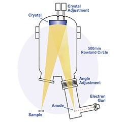
Image Credit: Thermo Fisher Scientific – Electron Microscopy Solutions
With a 500 mm Rowland circle and an Al anode (or dual Al and Ag anode if choosing the dual monochromator option), this twin-crystal, micro-focusing monochromator lets users choose any sample spot size between 200 µm and 900 µm.
Lens and Analyzer
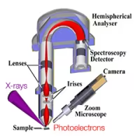
Image Credit: Thermo Fisher Scientific – Electron Microscopy Solutions
With a single analyzer path, the same instrument parameters (such as pass energy) can be used for spectroscopy and imaging, as well as the optimized lens and analyzer system on the ESCALAB QXi XPS Microprobe.
Ion Gun
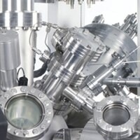
Image Credit: Thermo Fisher Scientific – Electron Microscopy Solutions
The ESCALAB QXi XPS Microprobe offers two alternatives for swift and high-resolution depth profiling. It includes the standard EX06 ion gun, optimized for monatomic ion sputtering and ion scattering spectroscopy. Additionally, it provides the option of the monatomic and gas cluster ion source, MAGCIS, which is versatile in performing monatomic ion profiling and cluster ion profiling. Both ion sources are able to be used to perform low energy ion scattering spectroscopy (ISS), which is a technique included as standard on the ESCALAB QXi.
Alignment and Calibration

Image Credit: Thermo Fisher Scientific – Electron Microscopy Solutions
To evaluate sensitivity, calibrating the ion source, aligning the X-Ray monochromator, adjusting the linearity of the analyzer energy scale, and figuring out the analyzer’s transmission function, a standards block is supplied. This contains standard reference samples of copper, silver, and gold, a phosphorescent sample for viewwing the X-ray beam, and apertures for ion beam alignment.
Sample Manipulator
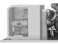
Image Credit: Thermo Fisher Scientific – Electron Microscopy Solutions
Accurate sample alignment for analysis is made possible by the computer-controlled, 5-axis high-precision translator (HPT). It can be used to automatically swap out sample holders and carry out queued experiments when combined with the recently developed automated sample loading system.
Preparation Chambers
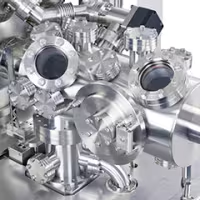
Image Credit: Thermo Fisher Scientific – Electron Microscopy Solutions
The Preploc chamber, a hybrid sample entry lock and preparation chamber, has ports that can hold various sample preparation tools, including gas admission, sample parking, ion guns, high-pressure gas cells, and heating/cooling probes.
Flood Electron Source
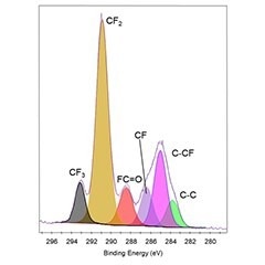
Image Credit: Thermo Fisher Scientific – Electron Microscopy Solutions
When analyzing non-conducting samples with the monochromatic X-Ray source, an electron source coaxial with the analyzer input lens is used for charge compensation in conjunction with a second flood source to supply ions at very low energy to ensure a near zero potential at the surface - eliminating the need for charge referencing in most cases. This assists with data interpretation by removing the uncertainty in the energy calibration when performing insulator analysis.
Detectors
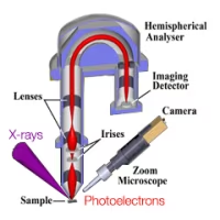
Image Credit: Thermo Fisher Scientific – Electron Microscopy Solutions
Two detector systems are installed on the ESCALAB QXi XPS Microprobe: one is designed for spectroscopy and consists of an array of six-channel electron multipliers, while the other is intended for parallel imaging and consists of two channel plates and a continuous position-sensitive detector. The two detector arrangement ensures that each system is optimised for it's function within the spectrometer.
Digital Control
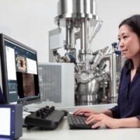
Image Credit: Thermo Fisher Scientific – Electron Microscopy Solutions
Since the Windows Software-based Avantage data system controls all analytical functions, the entire analysis process can be carried out remotely if necessary.
Sample Alignment
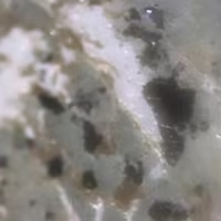
Image Credit: Thermo Fisher Scientific – Electron Microscopy Solutions
The Avantage Data System regulates every movement axis on the sample stage, and a high-definition digital video camera is attached to the device and precisely positioned in relation to the analysis position.
Vacuum System
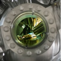
Image Credit: Thermo Fisher Scientific – Electron Microscopy Solutions
To optimize the effectiveness of the magnetic shielding, the analysis chamber is made of 5 mm-thick mu-metal. It is pumped with a turbomolecular pump and a titanium sublimation pump, enabling it to attain ultra-high vacuum of better than 5×10⁻¹⁰ mbar after bakeout.
Connectivity

Image Credit: Thermo Fisher Scientific – Electron Microscopy Solutions
For even quicker measurement area identification, measurement coordinates can be imported into Avantage Data System from specialized microscopy systems using Thermo Scientific Maps Software. By adding XPS spectroscopy and imaging data, surface chemistry and structural data can be directly compared with Maps Software.
Specifications
Source: Thermo Fisher Scientific – Electron Microscopy Solutions
| . |
. |
| Monochromated X-ray source |
- Micro-focused Al K-Alpha source or optional micro-focused dual-anode Al K-Alpha and Ag L-Alpha source
|
| Analyze |
- 180°, double-focusing, bi-polar hemispherical analyzer with dual detector system for spectroscopy and imaging
|
| Ion source |
- Monatomic EX06 ion source supplied as standard; option for MAGCIS dual mode ion source
|
| Vacuum system |
- Two turbo molecular pumps for entry and analysis chambers, with rotary pumps or oil-free backing pumps; auto-firing, 3-filament titanium sublimation pump for analysis chamber
|
| Sample stage |
- 5-axis sample stage with sample heating and cooling capabilities
- Optional fully automated sample exchange from load-lock parking post to analysis chamber
|
| Included standard analysis techniques |
- X-ray photoelectron spectroscopy (XPS)
- Reflected electron energy loss spectroscopy (REELS)
- Ion scattering spectroscopy (ISS)
|
| Optional analysis techniques |
- UV lamp for UV photoelectron spectroscopy (UPS)
- Non-monochromated X-ray source for HAXPES
- Field emission electron source for Auger electron spectroscopy (AES) and SEM
- Inverse photoemission spectroscopy (IPES)
- EDS detector
|
| Optional accessories |
- Glove box, vacuum transfer vessel, EX03 broad beam ion source, platter camera
|
| Sample preparation options |
- Heat/cool sample holder, additional preparation chamber, bakeable 3-gas admission manifold, fracture stage, sample parking facility
|