The Thermo Scientific Volumescope 2 Scanning Electron Microscope (SEM) is an advanced serial block-face imaging system. The device features user-friendly technology to maintain control over research and safeguard a variety of potential samples with tried-and-tested solutions for each stage of the acquisition process.
Block Face Imaging
Understanding the intricate three-dimensional structure of biological systems and soft materials is essential for understanding the relationship between structure and function. For automated, three-dimensional (3D) reconstruction of large tissue volumes, serial block-face SEM (SBF-SEM) combines in situ sectioning and imaging of plastic-embedded tissue blocks within the SEM vacuum chamber.
The minimum section thickness once limited axial resolution, but truly isotropic resolution is now possible with the addition of multi-energy deconvolution.
3D SEM
After the block face is sectioned in situ using a diamond knife, the newly exposed tissue is photographed numerous times at progressively higher accelerating voltages. These images are then used in a deconvolution algorithm to derive several optical subsurface layers, forming a 3D subset. By repeating this cycle, the Volumescope 2 SEM provides isotropic datasets with 10 nm Z-resolution.
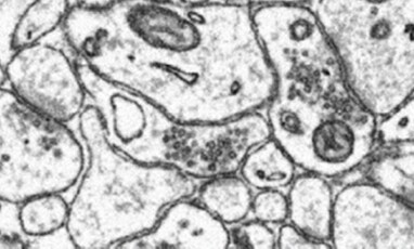
Mouse brain sample imaged on the Volumescope 2 SEM. Fine features such as vesicles and synaptic clefts can be observed. Image Credit: Dr Gabor Nyiri, Institute of Experimental Medicine, Budapest, Hungary
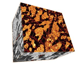
Volume reconstruction of a mouse heart muscle. Data acquired by a combination of physical and virtual slicing in high vacuum mode. Volume = 16×15×11 μm3 with a nominal resolution of 5×5×10 nm. Specimen courtesy: Dr Madesh Muniswamy, Temple University, USA. Image Credit: Thermo Fisher Scientific – Electron Microscopy Solutions
Polymer Microstructures
High-resolution imaging in 3D is imperative when trying to understand the properties and microstructure of polymer materials. The Volumescope 2 SEM combines in situ sectioning and imaging of polymers within the SEM vacuum chamber in a fully automated fashion to allow for the reconstruction of large volumes with truly isotropic 3D resolution.
Visualizing a sizable volume at these resolutions is essential to show how areas with various microstructures impact the material’s overall properties. To improve performance forecasts for new filter designs, the Volumescope 2 SEM, for instance, can recreate a large representative volume of a filtration membrane, providing accurate transport properties.
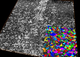
Serial block-face 3D imaging of a polymer blend sample. Here, the 3D volume reconstruction of the complete dataset (122 μm × 122 μm × 10 μm) is shown, with a segmented region inset revealing the particle distribution in the matrix. Sample courtesy of Dr. Armin Zankel (FELMI-ZFE Graz, Austria). Image Credit: Thermo Fisher Scientific – Electron Microscopy Solutions
Key Features
Easy-to-Use Technology
Easy-to-use technology allows users to stay in control of their experiments. Reuse jobs and system settings and select multiple regions of interest during job acquisition.
Protect Valuable Samples
Protect valuable samples with tested solutions at every acquisition step: debris trap and swipe features ensure sample quality; low vacuum detector enables imaging of highly charged samples.
Visualize and Navigate During the Acquisition
Visualize and navigate during acquisition with Thermo Scientific Amira Live Tracker Software to optimize/control your outcome/Automate large 3D volume acquisitions as well as reconstructions.
Easy and Fast Mounting Microtome Exchange
Easy and fast mounting microtome exchange for normal SEM operation or automated array tomography with the addition of Thermo Scientific Maps Software.
Specifications
Source: Thermo Fisher Scientific – Electron Microscopy Solutions
| . |
. |
| Electron optics |
- High-resolution field emission SEM column
- High-stability Schottky field emission gun
- Compound final lens: a combined electrostatic, field-free magnetic and immersion magnetic objective lens
- Dual objective lens combining electromagnetic and electrostatic lenses
|
| Source lifetime |
24 months
|
| Automation |
- Auto bakeout
- Auto start
- No mechanical alignments
- Automated heated apertures
- User guidance and column presets
|
| Stage |
Double stage scanning deflection
|
| Electron beam |
- Continuous beam current control and optimized aperture
- Beam current range: 1 pA to 400 nA
- In serial block-face imaging SEM: 50 pA – 3.2 nA
- Landing energy range: 20 eV – 30 keV
- Accelerating voltage range: 200 V – 30 kV
- In serial block-face imaging SEM: 500 V – 6 kV
- Resolution at optimum working distance and routine SEM high-vacuum imaging
- 0.8 nm at 30 keV STEM
- 0.7 nm at 15 keV
- 1.0 nm at 1 keV
|
| Chamber |
- Inside width: 340 mm
- Analytical working distance: 10 mm
- Ports: 12
|
| Detectors |
- Serial block-face imaging detectors
- T1 segmented lower in-lens detector
- VS-DBS: LoVac lens-mounted BSED
- T2 in-lens detector
- Everhart-Thornley SE detector (ETD)
- IR-CCD
- Low-vacuum SE detector
- STEM 3+ – Retractable segmented detector
- Additional detectors available
|
| Vacuum system |
- Complete oil-free vacuum system
- 1 × 220 l/s TMP
- 1 × PVP-scroll
- 2 × IGP
- Chamber vacuum (high-vacuum) <6.3 × 10⁻⁶ mbar (after 72 hours pumping)
- Low-vacuum mode up to 50 Pa for charge compensation of non-conductive samples
- Evacuation time: ≤3.5 minutes
|
| Sample holders |
- Standard multi-purpose holder; uniquely mounts directly onto the stage; hosts up to 18 standard stubs (Ø12 mm), three pre-tilted stubs, two vertical and two pre-tilted row-bar holders
- Each optional row-bar accommodates six S/TEM grids
- Wafer and custom holders
|